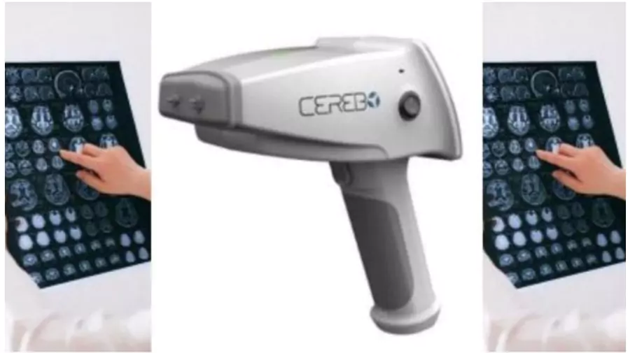
The Indian Council of Medical Research (ICMR), in collaboration with AIIMS Bhopal, NIMHANS Bengaluru, MDMS Secretariat, and Bioscan Research, has developed CEREBO, a portable diagnostic tool for Traumatic Brain Injuries (TBI).

About Traumatic Brain Injuries (TBI)
- Cause: TBI is caused by sudden trauma or injury to the head, disrupting normal brain function.
- Severity: It ranges from mild concussions to severe, life-threatening injuries.
- Major causes in India:
-
- Road Traffic Accidents – 60%.
- Falls – 20–25%.
- Violence – ~10%.
- Consequences: Bleeding, swelling, cognitive impairments, behavioural changes, physical disabilities, and long-term neurodegenerative risks.
About CEREBO
- Nature: Handheld, portable, and non-invasive diagnostic device for traumatic brain injuries (TBIs).
- Technology: Built on near-infrared spectroscopy combined with machine learning.
- Speed: Detects intracranial bleeding and edema in under one minute.
- Output: Provides colour-coded, radiation-free, and cost-effective diagnostic results.
- Safety: Suitable for infants and pregnant women.
- Ease of Use: Can be operated by paramedics or unskilled personnel with just 30 minutes of training.
- Applications: Designed for ambulances, trauma centres, rural clinics, and disaster response units.
Significance of CEREBO
- Early Detection: Helps in timely diagnosis where CT (Computed Tomography) and MRI (Magnetic Resonance Imaging) facilities are unavailable or delayed.
- Accessibility: Can be widely used in rural and underserved areas lacking advanced infrastructure.
- Efficiency: Reduces time-to-decision in emergencies, optimises triage, and lowers imaging costs.
- Global relevance: Potential adoption in military healthcare and disaster response systems.
- Primary-Care Utility: Facilitates rapid, reliable triage at primary-care centres and ambulances, improving survival rates.
- Resource-Limited Settings: Effective during accidents, falls, and violence-related injuries, which are common in rural regions.
Need for the CEREBO Device
- High Burden of TBI in India:
- Over 100,000 deaths annually due to head injuries.
- More than 1 million people suffer serious brain injuries each year.
- Limitations of existing methods:
- Glasgow Coma Scale – prone to error and subjectivity.
- CT/MRI – require costly infrastructure, trained specialists, and are often inaccessible in rural areas.
- Criticality of early detection:
- Half of TBI deaths occur within the first two hours of injury.
- Secondary brain injury (post-impact swelling/bleeding) worsens outcomes.
- Early diagnosis improves mortality and disability outcomes.
Feature |
CT (Computed Tomography) |
MRI (Magnetic Resonance Imaging) |
Technology |
Uses X-rays with computer processing to create cross-sectional images. |
Uses strong magnetic fields and radio waves to generate detailed images. |
Best For (Use Cases) |
- Head injuries & skull fractures
- Detecting bleeding & clots
- Chest, abdomen, and bone injuries
- Fast diagnosis in emergencies/accidents
|
- Brain and spinal cord injuries
- Tumors & soft tissue evaluation
- Joint, ligament, and cartilage injuries
- Stroke and neurological disorders
|
Speed |
Very fast (a few minutes) – useful in emergencies. |
Slower (30–60 minutes per scan). |
Image Detail |
Better for bones and acute bleeding. |
Superior for soft tissue, nerves, and brain structures. |
Cost |
Cheaper than MRI. |
More expensive. |
Risks |
- Radiation exposure (due to X-rays)
- Not ideal for repeated scans.
|
- No radiation, but not suitable for patients with metal implants/pacemakers
- It can cause claustrophobia in some patients.
|