A new tool to detect viral infections in cells using light has been developed by researchers. The tool has been developed by researchers from Harvard University, Cambridge, and Jiangsu University, Zhenjian.
- Published In: The paper was published in the journal Science Advances.
Viruses
They are microscopic organisms that can cause infection to a variety of hosts ie. Humans, plants, animals, bacteria and Fungi.
- Structure: They’re a small piece of genetic information (DNA or RNA) inside of a protective shell (capsid). Some viruses also have an envelope. Viruses can’t reproduce without a host.
- They’ll survive outside of a host until their capsid breaks down over time.
- Reproduction: Viruses aren’t made up of cells, therefore they generally can’t make copies of themselves on its own. Instead, they carry instructions with them and use a host cell’s equipment to make more copies of the virus.
- Types: Influenza viruses, Human herpesviruses, Coronavirus, Human papillomaviruses, Enteroviruses, Flaviviruses, Orthopoxviruses, Hepatitis viruses.
- Diseases: Viruses can cause flu, the common cold and COVID-19.
|
New Light-Based Tool Detects Viral Infections – Study
- Objective: To develop a tool to identify the fingerprints of cellular viral infection in a body for early detection.
Enroll now for UPSC Online Course
- Experiment:
- Sample: Researchers infected cells from a pig’s testicles with pseudorabies virus, shone light on them through a microscope, and tracked how changes in the cells distorted the light.
- A viral infection can stress cells and change their shapes, sizes, and features with the changes becoming more pronounced as the infection spreads and the body becomes diseased.
- These distortions were recorded at different points of time so that the light data mimicked a progressing viral infection.
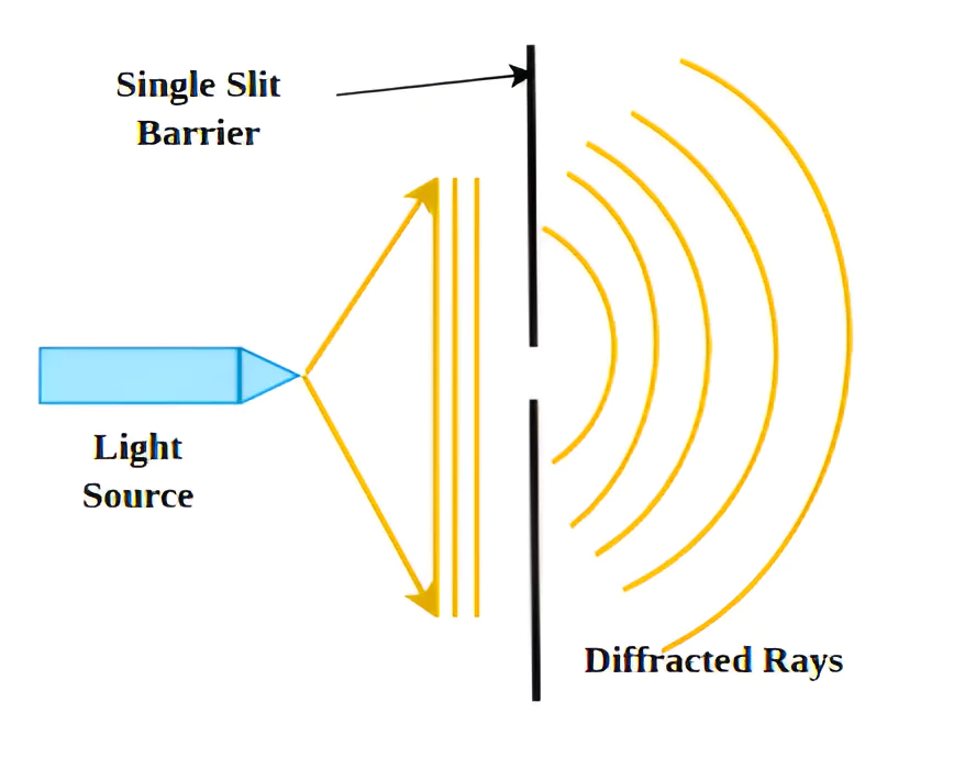
- Distortion of Light: This distortion was due to the pattern made by the Diffraction Process of alternating light and dark rings or stripes around a dark centre once it reaches, say, a wall.
- Comparison: These distortions of light were compared between the infected and the healthy cells and the difference reported between the two light patterns.
- Fingerprint of virus-infected cells: The cellular changes were then translated into patterns that could be used to say if a cell had been infected.
- Parameters: The fingerprint was based on two parameters,
- The contrast between the light and dark stripes
- The inverse differential moment (a mathematical value defining how textured the diffraction pattern was)
- Finding: The method can differentiate between uninfected, virus-infected, and dead cells.
- Virus-infected cells: They were elongated and had more clear boundaries than uninfected cells, changing the contrast between light and dark stripes of the diffraction fingerprint, and increasing the differences in light intensity.
Advantages of New Light-Based Tool Detects Viral Infections
- Accuracy: The light based method was found to detect viral infections as accurately or even more accurately than the standard method.
- Cheap: The new method is cheaper with the cost being one tenth of the standard method whose equipment cost using chemical reagents is about Rs 2.5 lakh
- Independent of supply chain issues: Many research facilities around the world procure reagents from other places, adding potential time delays and vulnerability of their research to supply-chain inefficiencies.
- Fast: The new method reportedly takes only about two hours to detect virus infected cells, against the 40 hours the current standard required.
- Livestock management: The new tool can help spot viral infections in the bodies of livestock as well as for the selection and breeding of excellent livestock and poultry species at the cellular level.
- Early detection: The new method could help catch viral infections early and can help detect viral infections in general helping the stakeholders take preventive measures in time to avoid significant losses.
- Identify a new Virus: The tool’s generic nature could also be an advantage by catching a viral infection that is not due to existing pathogens, perhaps even a new virus.
- Surveillance: A rapid and cost-effective way to detect viral infections could help improve surveillance and reduce the cost of selecting healthy animals or birds for breeding.
- The existing methods to select animals for breeding require expensive DNA-sequencing tools.
- Capacity building: The light based tool could also help low- and middle income countries to realize the WHO’s recommendation of, rapid detection, reporting and responding to animal outbreaks as the first line of defence” against the spread of viruses.
Standard method to detect Viral Infections in cells
- Chemical Reagents: A Researcher isolates infected cells in the lab and add chemical reagents like dimethyl thiazolyl, diphenyl tetrazolium bromide to them.
- The enzymes in the cells ie. (oxidoreductases and dehydrogenases), react with the reagent to produce purple crystals of a chemical entity called formazan. The reagent destroys the cell afterward.
- Color change: The color change is the indicator whether cells could have had a viral infection. Cells dying of a viral infection lack these enzymes and thus produce little to no amounts of formazan crystals.
|
Diffraction

It is the spreading of waves around an obstacles.
- Diffraction takes place with sound; with electromagnetic radiation ( light, X-rays, and gamma rays) and with very small moving particles such as atoms, neutrons, and electrons, which show wavelike properties.
- The phenomenon: It is the result of interference (i.e., when waves are superimposed, they may reinforce or cancel each other out) and is most pronounced when the wavelength of the radiation is comparable to the linear dimensions of the obstacle.
- Consequence: A consequence of diffraction is that sharp shadows are not produced.
|
Enroll now for UPSC Online Classes
![]() 30 May 2024
30 May 2024