What is Cryo-Electron Microscopy (cryo-EM)?

10 Jun 2025

What is Cryo-Electron Microscopy (cryo-EM)?

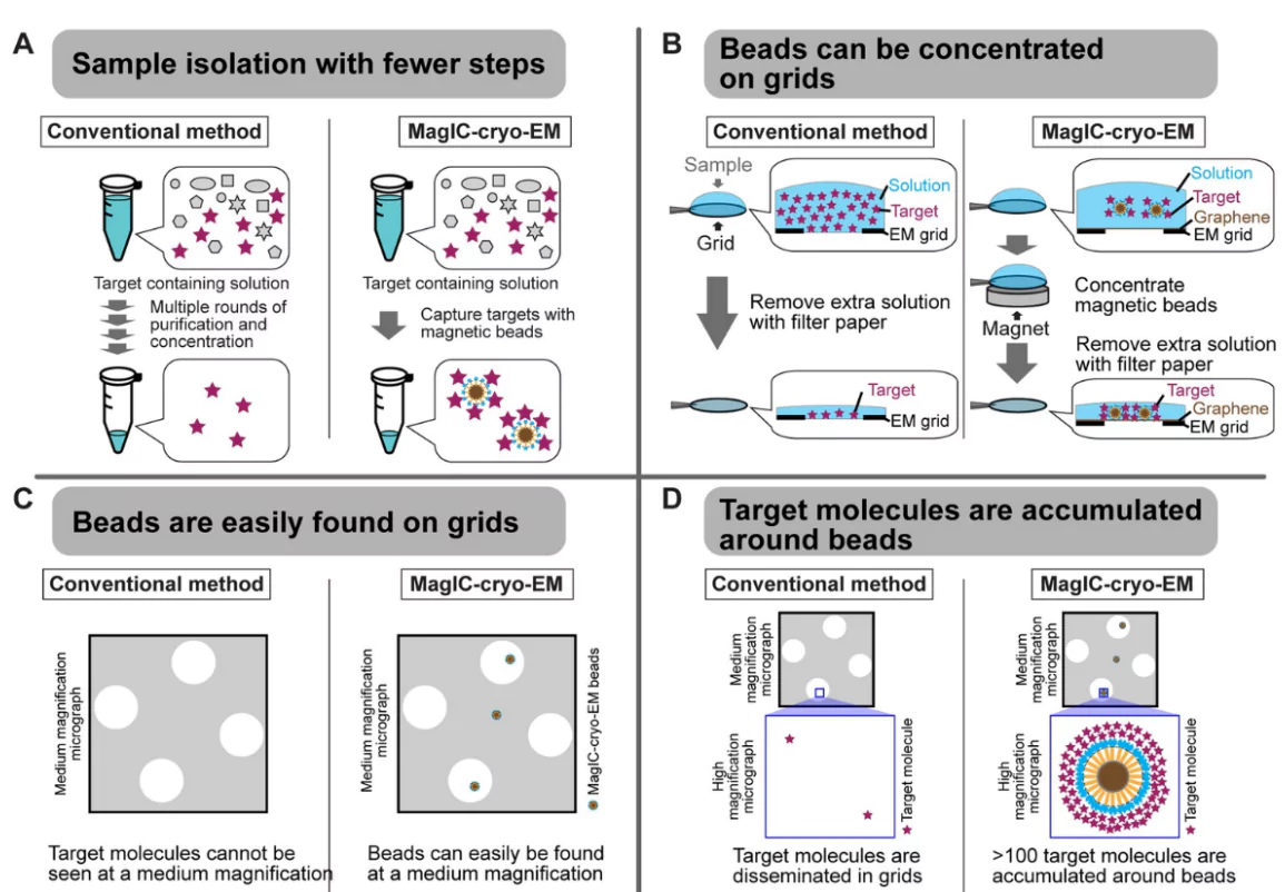
U.S. researchers have developed a new cryo-EM method called MagIC (Magnetic Isolation and Concentration), allowing imaging of biological molecules at concentrations 100 times lower than before.

About Cryo-Electron Microscopy

  • Cryo-electron microscopy (cryo-EM) is an advanced imaging technique used to study the detailed structure of biological molecules like proteins, viruses, or DNA at very high resolution.
  • Core Principle: Biological samples are rapidly frozen in vitreous (non-crystalline) ice and then imaged using an electron beam under cryogenic conditions. 
    • This preserves their natural structure and prevents radiation damage.
  • Limitation: Cryo-electron microscopy (cryo-EM) requires high concentrations of biological molecules. 
    • Rare or hard-to-isolate molecules are difficult to image due to low availability.
  • National Cryo EM Facilities: The first national cryo-EM facility was established at National Centre for Biological Sciences (NCBS)in 2017 and then subsequently in IISc, Bangaluru, and RCB Faridabad, IIT Kanpur.

Key Features Of MagIC (Magnetic Isolation and Concentration)

  • Magnetic Clustering: The new method works by attaching molecules of interest in a sample to 50-nm beads, then using a magnet to clump the beads together.
  • Efficiency at Low Concentration: This technique allows imaging even with sample concentrations as low as 0.0005 mg/ml, significantly reducing sample requirements.
  • Accelerated Microscopy: Beads are easily visible even at low magnification, enabling rapid identification of particle-rich regions and faster data acquisition.
  • Noise Reduction Strategy i.e. DuSTER: Small particles often hide in background noise
    • To pull them out, the researchers built a computer workflow called Duplicated Selection To Exclude Rubbish (DuSTER)
    • Two-Step Validation: The DuSTER algorithm picked each particle twice, kept those that landed in the same place after two rounds of 2D or 3D classification, and threw the rest away.
    • Even when cryo-electron microscope (cryo-EM) images are full of noise and look unusable, the DuSTER software is able to identify and extract the useful, high-quality particle images from them.

Cryo-Electron Microscopy

Significance of MagIC

  • Scientific Advancement: Allows structural study of rare, low-concentration biomolecules.
  • Sample Efficiency: Lowers the sample demand to just 5 nanograms per grid
  • Data Quality: Combines magnetic targeting with computer filtering for improved accuracy.
  • Speed: The scientists can quickly move the microscope to regions rich in particles, speeding up data collection.
What is Cryo-Electron Microscopy (cryo-EM)?

Explore UPSC Foundation Course

Need help preparing for UPSC or State PSCs?

Connect with our experts to get free counselling & start preparing

Aiming for UPSC?

Download Our App

      
Quick Revise Now !
AVAILABLE FOR DOWNLOAD SOON
UDAAN PRELIMS WALLAH
Comprehensive coverage with a concise format
Integration of PYQ within the booklet
Designed as per recent trends of Prelims questions
हिंदी में भी उपलब्ध
Quick Revise Now !
UDAAN PRELIMS WALLAH
Comprehensive coverage with a concise format
Integration of PYQ within the booklet
Designed as per recent trends of Prelims questions
हिंदी में भी उपलब्ध

<div class="new-fform">


    </div>

    Subscribe our Newsletter
    Sign up now for our exclusive newsletter and be the first to know about our latest Initiatives, Quality Content, and much more.
    *Promise! We won't spam you.
    Yes! I want to Subscribe.